MRI Spots Progression of Facioscapulohumeral Muscular Dystrophy Better Than Standard Methods, Study Reports


MRIs can pick up subtle muscle changes that standard tests cannot in slowly progressing disorders such as facioscapulohumeral muscular dystrophy, or FSHD, a study reports.
The research in the journal PLoS ONE was titled “Long-term follow-up of MRI changes in thigh muscles of patients with Facioscapulohumeral dystrophy: A quantitative study.”
FSHD is a muscular disorder that primarily affects face, shoulder, and arm muscles, but also lower-extremity and pelvic muscles.
Doctors have difficulty evaluating FSHD muscle changes and the disease’s progression because current testing methods are unable to detect small differences occurring over short periods. Making it more challenging is that patients who are motivated can appear to have better muscle performance than they actually do.
This means that doctors need better ways to evaluate slowly progressing diseases such as FSHD.
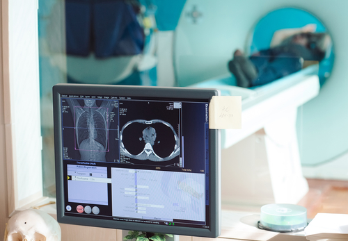
Physicians use magnetic resonance imaging, or MRI, to assess muscle changes in several disorders, including FSHD. Studies have shown that imaging-based evaluation of muscles can detect disease progression better than standard methods.
Despite these findings, MRI has yet to supplant standard methods as the main yardstick for measuring the progression of slowly developing diseases.
A research team at Aix Marseille University in France decided to use both standard methods and MRI to evaluate muscle changes in 35 FSHD patients and 15 age-matched healthy volunteers. The standard methods included testing muscles manually and measuring patients’ movement abilities.
The imaging detection method the team used was looking for changes in MRI pixel intensity distribution. It spotted differences in muscle loss and fat infiltration in thigh muscles that could translate into disease progression.
Researchers’ final task was to correlate the results of the standard and MRI tests with patient outcomes.
The team found a strong correlation between the standard and MRI readings of patients’ muscle strength when the study started.
Standard methods failed to detect muscle change during the one to two years of follow-up, but imaging did. While standard methods generated a finding that no muscle deterioration had occurred, MRIs indicated that it had decreased by 2.3 percent a year.
The difference was that imaging detected an increase in fat and a decrease in muscle mass that standard methods were unable to spot.
“This slow disease progression and the poor sensitivity of the commonly used clinical scores mandates the utilization of more sensitive biomarkers which could be used to appropriately assess disease progression and the potential effects of therapeutic interventions,” the researchers wrote.
Overall, they concluded that MRI “may reveal muscle changes which cannot be evidenced using clinical scales and therefore may provide reliable and non-invasive outcome measures for clinical trials” of therapies.