
Key mechanism found for muscle stem cells survival after transplant
Implications 'immense' for treating MD, other diseases, researcher says
Written by |

The expression of a protein called ACTC1 in muscle fibers is critical for muscle repair — and vital to the survival of transplanted human muscle stem cells — a new study shows.
These findings may have important implications for the development of cell therapies for muscular dystrophy and other disorders that affect the muscles.
“With our discovery, the development of ‘muscle in a dish’ is one step closer to reality. We’ve been researching this for years, and [its] implication for treating disease and muscle disorders and tears are immense,” Michael H. Hicks, PhD, lead author of the study at the University of California, Irvine (UCI), said in a university press release.
The team’s work to develop muscle stem cells in the lab is now under review for a patent in the U.S., as well as in Europe and Japan, the release stated.
The study, “Regenerating human skeletal muscle forms an emerging niche in vivo to support PAX7 cells,” was published in Nature Cell Biology.

Looking for solutions to a technical challenge in cell therapies
Stem cells are specialized cells that are able to grow and differentiate to form other types of cells. Within muscle tissue, there are specialized cells of this type that normally are able to make new muscle fibers when muscle is damaged. In muscular dystrophies however, muscle damage occurs faster than these stem cells are able to repair it.
Theoretically, treatments that could transplant healthy muscle stem cells into a patients’ muscles could be a useful treatment strategy for muscular dystrophies. Such a strategy might allow production of new healthy muscle fibers.
However, getting muscle stem cells to actually stick around and make new healthy muscle tissue after being transplanted has proven a technical challenge.
Now, researchers uncovered new insights into the biology of these specific stem cells that may help to advance this treatment strategy.
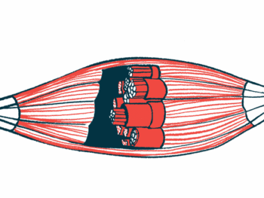
Muscle stem cells are marked by the expression of a protein called PAX7, and they are normally found at certain physical locations or compartments in muscle tissue, referred to as niches.
Working in a mouse model of Duchenne muscular dystrophy, the scientists showed that transplanted human muscle stem cells are able to grow to form new muscle fibers. But these human cells were almost entirely segregated by themselves, forming muscle fibers of only human cells. The transplanted human stem cells were almost exclusively located in the niches of these human muscle fibers, rather than taking up residence in the mice’s existing muscle niches.
At first, the researchers thought this difference might be because the mice’s existing stem cells were not leaving enough room for transplanted human cells to enter. But even when mice were engineered to lack their own muscle stem cells, the transplanted human cells still stuck to human-only niches.
This finding implies that there are specific biological signals that the stem cells receive in the human niches that are absent in the mouse niches. The researchers noted that this finding with these stem cells differs from the behavior of other types of stem cells, such as blood stem cells.
This work has global implications for cell therapies and provides a new model system to study how to support human regeneration and niche emergence.
In a further series of experiments, the researchers found that the human muscle fibers created by transplanted cells in the mice expressed many proteins that are normally present during the early development of muscles in the womb. In particular, the muscle fibers surrounding the niche expressed high levels of the protein ACTC1.
The scientists showed that, when the muscle fibers were modified to lack ACTC1, far fewer stem cells were able to survive in the niche — and when the muscle stem cells were engineered to lack the characteristic marker PAX7, the muscle fibers expressed less ACTC1, suggesting these two cell types communicate with each other to support the muscle stem cell niche.
“We tailored a high-dimensional spatial analysis platform to identify how transplanted human progenitor cells and myofibers [muscle fibers] in a mouse were communicating,” said Ben Clock, a graduate student at UCI and study co-author.
The identification of these systems, which support muscle stem cells in the body, will be key to advancing treatments for muscle disorders, including in muscular dystrophy, according to the researchers.
“This work has global implications for cell therapies and provides a new model system to study how to support human regeneration and niche emergence,” the team concluded.
Leave a comment
Fill in the required fields to post. Your email address will not be published.