Study Finds That Muscle Fibrosis Inhibition Impacts in Duchenne Muscular Dystrophy


In a recent study published in Oncotarget, researchers found an inverse correlation between the level of muscle fibrosis, utrophin and the number of revertant myofibers in Duchenne Muscular Dystrophy (DMD).
Results from this study reveal common links between the fibrotic and utrophin-synthesis pathways and offer new insights into the regulation of utrophin synthesis in Duchenne Muscular Dystrophy.
The most common form of X-linked muscle dystrophy (MD) is Duchenne Muscular Dystrophy (DMD), which affects 1 in 3,500 live males at birth. Progress of muscle degeneration and worsening clinical symptoms leads to death in the late teens or early twenties as a result of respiratory or cardiac failure.
DMD is characterized by absence of a protein named dystrophin in skeletal muscles. The dystrophin-glycoprotein complex (DGC) connects the actin cytoskeleton of myofibers to the extracellular matrix (ECM) and is essential to the contractile structure of muscle.
The preliminary stage of the disease is characterized by the presence of focal groups of necrotic myofibers, muscle hypertrophy, and abnormally high levels of muscle creatine kinase (CK).
The increase in collagen deposition in DMD initiates a vicious cycle in which further muscle destruction leads to joint contractures, loss of ambulation, and death from respiratory or cardiac failure. Although DMD is caused by frame-disrupting mutations in the DMD gene that prevent the full translation of dystrophin in muscles of DMD patients and in mdx mice — the murine model of DMD – both exhibit sporadic low percentages of dystrophin-positive myofibers known as revertant fibers (RFs).
In the study titled “Inhibition of muscle fibrosis results in increases in both utrophin levels and the number of revertant myofibers in Duchenne muscular dystrophy,” Mark Pines from the Institute of Animal Sciences, Volcani Center, Bet Dagan in Israel and colleagues found in patient quadricep femur biopsies (n = 6, ages between 3–9 years) an inverse correlation between the levels of collagen type I – representing fibrosis — and the levels of utrophin.
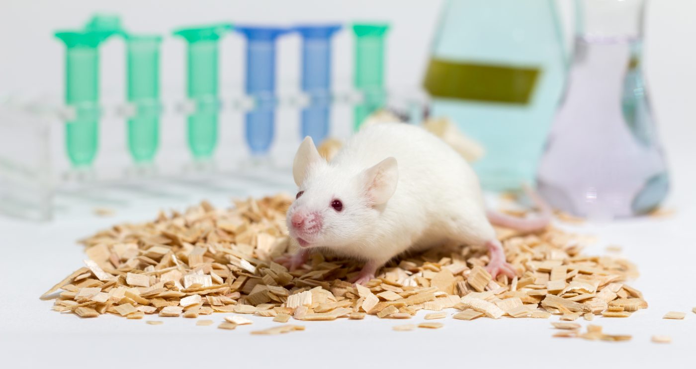
In the mice models (n = 6/group), inhibition of fibrosis by halofuginone resulted in increases in the levels of utrophin.
The researchers also found that the utrophin/fibrosis relationships were not limited to collagen type I, but also applied to other constituents of the fibrosis machinery.
Results also revealed that inhibition of collagen type I levels was associated with an increase in the numbers of revertant myofibers, both as single myofibers and in clusters in the diaphragm and the gastrocnemius.
In the study, the researchers indicate that because the existing strategies to cure MDs, such as gene and cell-replacement therapies have met with some difficulties, the development of complementary and supportive therapies that slow disease progression and improve patients’ quality of life is critically important.
According to the researchers, a more realistic goal for the near future would be to treat the main pathological mechanisms, e.g., by inhibiting muscle fibrosis and simultaneously increasing utrophin levels and RF numbers in the dystrophic muscle, to delay the progressive loss of muscle strength, and improve muscle integrity and function.