
Abdominal Muscles Involved in Cardiopulmonary Pathology in Subjects with Muscular Dystrophy
Written by |
 Researchers at The University of Chicago recently revealed that the abdominal muscles can reflect the cardiopulmonary pathology status in muscular dystrophy mouse models. The study was published in the Journal of Neuromuscular Diseases and is entitled “Cardiac function in muscular dystrophy associates with abdominal muscle pathology.”
Muscular dystrophy is characterized by a progressive skeletal muscle weakness that leads to the death and loss of muscle cells and tissue, compromising locomotion. It can also affect specific muscles involved in respiratory function, leading to breathing complications and cardiac problems. Various groups of muscles are known to be differently targeted in muscular dystrophies.
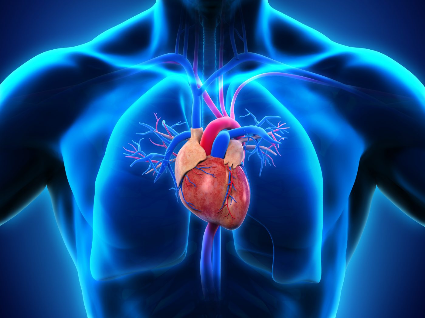
It is known that there is interdependence between cardiac and respiratory function in the body. In muscular dystrophy, where both cardiac and respiratory muscles can be affected, this interdependence between the two systems can accelerate pathology and disease progression.
[adrotate group=”3″]
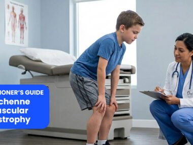
Researchers studied two mouse models of muscular dystrophy, namely, the mdx mouse model of Duchenne muscular dystrophy and the Sgcg mouse model of limb girdle muscular dystrophy, to assess the disease’s impact on different muscle groups and on cardiac function. Both mouse models exhibit a similar pathology to the one seen in human muscular dystrophy, including significant damage to the diaphragm muscle.
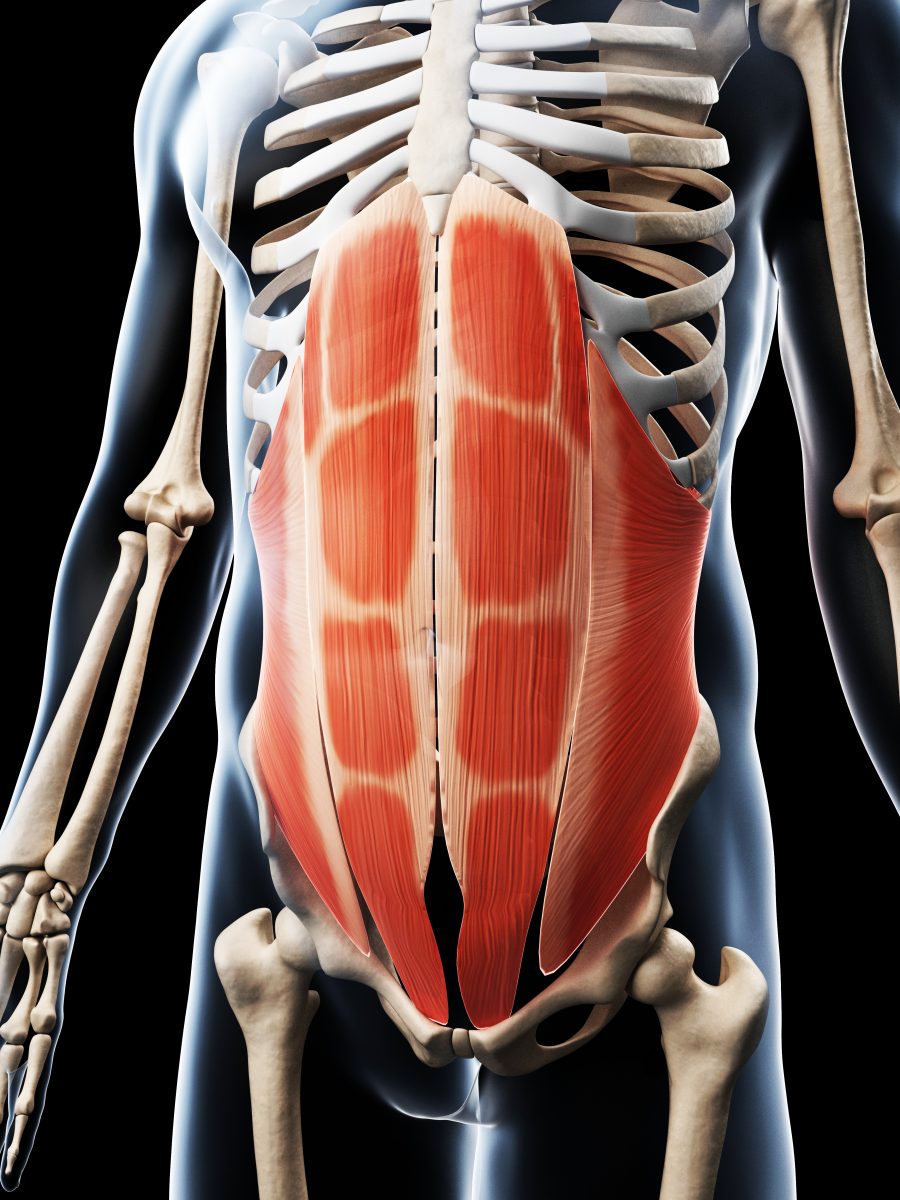
The team found that abdominal muscles can be affected by the disease, and that these muscles play an important role in respiratory support, especially when the diaphragm muscle has been damaged as a consequence of the disorder, suggesting that additional damage to the abdominal muscles can seriously compromise the respiratory function. In addition, abdominal muscles were found to have more fibrosis (scarred tissue) than other muscle groups. The degree of fibrosis in the diaphragm muscle was found to positively correlate with fibrosis in the heart’s left ventricle. On the other hand, abdominal muscle fibrosis was associated with an impaired left ventricular function, while it negatively correlated with fibrosis in the right ventricle and the diaphragm, suggesting that abdominal muscles seem to reflect the cardiopulmonary pathology of the disease.
It was concluded that the abdominal muscles are recruited as respiratory muscles in a muscular dystrophy situation, in agreement with data from human patients with this disease, and that this group of muscles could be studied further as disease biomarkers that reflect the cardiopulmonary pathology status.
“Supporting and maintaining proper cardiopulmonary function in neuromuscular disease is a mainstay of therapy. Maintaining diaphragm health has been the focus of many studies in both humans and mice with muscular dystrophy, but few studies have focused on supporting and evaluating the accessory muscles of respiration such as the abdominal muscles. Therapies that spare or protect the muscles of respiration in muscular dystrophy have been shown to slow down overall disease progression and prolong life. The accessory muscles of respiration, whether in human patients or animal models, may prove a viable target especially for therapy directed at specific muscle groups,” concluded the study’s senior author Dr. Elizabeth M. McNally in a news release.

Leave a comment
Fill in the required fields to post. Your email address will not be published.