Dystrophin Protein Essential For Maturation of Brain Cells, Study Finds

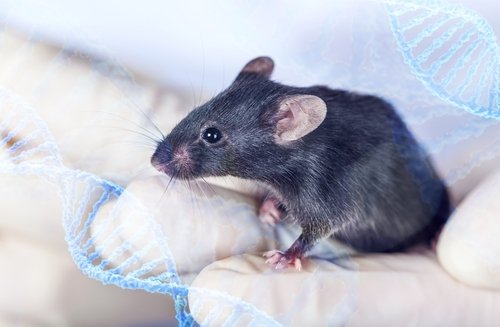
Researchers, using a mouse model of Duchenne muscular dystrophy (DMD), have discovered that a specific protein, dystrophin, plays an essential role in myelination during postnatal brain development.
The protein dystrophin plays a major role in DMD. Mutations in the dystrophin gene are a major contributing factor for the development of DMD. Dystrophin functions as a complex of related proteins, called the dystrophin-associated glycoprotein complex (DGC). This complex connects signaling molecules outside the cell to signalling proteins within the cell. In this way, the DGC plays a regulatory role in mediating cellular functions. Although DMD is largely characterized by muscle degeneration, many patients also suffer from neurodevelopment problems. The factors leading to neurodevelopment deficits are not well understood.
In this study, “Myelination is delayed during postnatal brain development in the mdx mouse model of Duchenne muscular dystrophy,” published in BMC Neuroscience, the authors explore the role of the dystrophin protein in the central nervous system.
Myelination is the process where a protective coating, myelin, is produced on the outside of specific brain cells. This process begins early in life, but is not completed until adolescence. In the brain, this function is performed by cells called oligodendrocytes. Maturation of oligodendrocytes is essential for their myelination function.
For the first time, the authors found that oligodendrocytes can express three different forms of dystrophin, Dp427, Dp140, and Dp71. In a mouse model, these different forms of dystrophin are expressed during different stages of development. Dp427 and Dp140 are predominant in immature oligodendrocytes, whereas Dp71 is the major form of dystrophin expressed in mature oligodendrocytes.
The mdx mice is the best characterized model of DMD. In this model, expression of the largest form of dystrophin, Dp427, is lost, which also is commonly observed in DMD patients. And similar to DMD patients, mdx mice also have abnormal brain function.
Using this mouse model, researchers observed changes in myelination during postnatal development. Compared to controls, in mice which lacked the Dp427 dystrophin protein, myelination occurred at later points during development. This effect was most significant in the cerebral cortex.
When looking at the postnatal maturation of oligodendrocytes, the researchers saw an accumulation of immature oligodendrocytes, or the oligodendrocyte progenitor cells (OPCs), in mdx mice that lacked a form of dystrophin compared to controls. Further studies showed that these OPCs failed to mature into fully functioning and myelinating oligodendrocytes.
To validate that loss of dystrophin is responsible for the defects observed in oligodendrocyte maturation and myelination, expression of all forms of dystrophin was decreased in oligodendrocytes in vitro. Similarly to the mdx mouse model, loss of dystrophin results in delayed maturation of oligodendrocytes.
These data suggest that proper expression of dystrophin is essential for maturation of oligodendrocytes and effective myelination of brain cells during development. The delayed myelination observed in mouse models will likely negatively affect the speed and coordinated timing that is needed for complex brain processing. This may explain some of the neurological deficits seen in patients with DMD.
The authors concluded that “loss of oligodendroglial dystrophin(s) may be a contributing factor in DMD-related neurological deficits.”


