EGF Protein Preserves Muscle Strength, Promotes Muscle Regeneration in DMD Mice, Study Shows
Written by |

A protein called epidermal growth factor (EGF) can help preserve muscle strength and increase muscle regeneration in a mouse model of Duchenne muscular dystrophy (DMD), a finding that may pave the way for new treatment strategies for DMD, researchers said.
The study, “EGFR-Aurka Signaling Rescues Polarity and Regeneration Defects in Dystrophin-Deficient Muscle Stem Cells by Increasing Asymmetric Divisions,” was published in the journal Cell Stem Cell.
Researchers from The Ottawa Hospital and the University of Ottawa in Canada had described in previous studies that, besides damage in muscle fibers, deficiency of the dystrophin protein in DMD also could disrupt the multiplication process of muscle stem cells. This would impair the ability of muscles to regenerate, leading to loss of skeletal muscle.
Muscle stem cells can multiply through a process called asymmetric division, which typically generates a copy of the stem cell with self-renewal ability and another cell that differentiates into a more specialized cell type.
To better understand how this division process was regulated, the team now conducted a screen in which they exposed muscle stem cells to 640 well-characterized pharmacological compounds.
They found that in the presence of inhibitors that blocked the activity of the EGF receptor, or EGFR, and Aurora kinase A (Aurka), the stem cells were unable to undergo asymmetric division. Specifically, upon several lab experiments, they found that EGFR activation recruits Aurka to induce asymmetric division.
Suppressing Aurka with small interfering RNAs (siRNAs), which can prevent the normal availability of the enzyme, impaired this type of stem cell division. In turn, blocking EGFR with lapatinib (marketed as Tykerb by Novartis as treatment for breast cancer) shifted asymmetric to symmetric divisions, which means the generation of two differentiated cells or two stem cells.
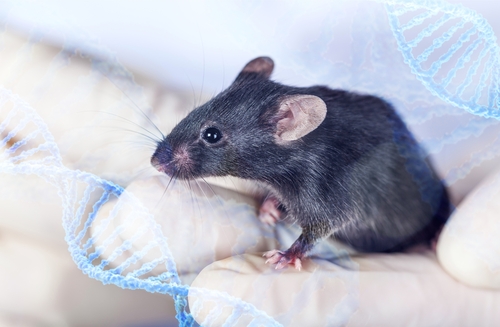
Next, the team genetically manipulated DMD mice by inserting a gene producing human EGF (the protein that binds to and activates EGFR) into the muscle during the onset of muscle degeneration. Long-term production of EGF led to 18% more muscle mass, 30% more muscle fibers, 32% greater muscle strength, and a marked reduction in muscle fibrosis (scarring), compared to non-EGF exposed mice. These benefits were found to be sustained for at least five months.
These findings were in line with those in muscle fibers collected from these mice, where increased levels of EGF led to a significantly higher rate of asymmetric division and the higher number of muscle cells.
When they directly injected a manmade version of EGF into mice muscles, researchers found increased levels of muscle stem cells, further suggesting that EGF could promote muscle regeneration.
“These results indicate that EGF treatment provides long-term enhancement of muscle strength in [humanized DMD] mice,” researchers wrote. “We envision that stimulation of muscle stem cell function can be combined with dystrophin restoration to restore muscle function in DMD,” they added.
As a next step, the team will try to identify a molecule that combines the same muscle effects of EGF upon delivery into the bloodstream. They also will assess the long-term effects of triggering the EGFR signaling pathway, as a potential therapy likely would be taken throughout life.
“This is a huge step forward in developing a new approach for treating [DMD],” Michael Rudnicki, PhD, said in a press release. Rudnicki directs the Regenerative Medicine Program and Sprott Centre for Stem Cell Research at Ottawa Hospital Research Institute, and is senior author of the study, “We were able to preserve the muscle strength and function that are usually lost over time in this disease,” he said.
“[DMD] is complex, and we will probably need a combination of treatments to address all aspects of the disease,” said Rudnicki, who also is a professor at the University of Ottawa.


