Immune Cells May Help Treat Muscular Dystrophy, UCSF-Led Team Finds
Written by |
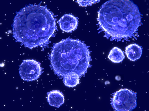
 An international research team led by University of California at San Francisco scientists has discovered that regulatory T cells (Tregs), a specialized subset of immune cells, suppress inflammation and muscle injury in a mouse model of Duchenne muscular dystrophy (DMD).
The scientists say Tregs have potential as therapeutic agents for DMD, an inherited disease that strikes children — almost always boys — leading to progressive muscle degeneration and early death.
The findings were published online October 15, 2014, in the American Association for the Advancement of Science journal Science Translational Medicine, entitled “Regulatory T cells suppress muscle inflammation and injury in muscular dystrophy“ (Sci. Transl. Med. 15 October 2014: Vol. 6, Issue 258, p. 258ra142 DOI: 10.1126/scitranslmed.3009925), coauthored by S. Armando Villalta and Jeffrey A. Bluestone of the Diabetes Center, University of California, San Francisco. Dr. Bluestone is also associated with the Departments of Pathology and Medicine, UCSF.
 Additional co-authors of the study are Marta Margeta, MD, PhD, assistant professor of pathology; from UCLA; Wendy Rosenthal, research associate, Amanjot Kaur, lab assistant and James G. Tidball, PhD, chair of molecular, cellular and integrative physiology from UCSF; Melissa J. Spencer, PhD, co-director of the Center for Duchenne Muscular Dystrophy; and Leonel Martinez, lab assistant; and Tim Sparwasser, MD, director of the Institute of Infection Immunology, Hannover, Germany.
The researchers examined the hypothesis that regulatory T cells (Tregs) modulate muscle injury and inflammation in the mdx mouse model of Duchenne muscular dystrophy (DMD). They explain that although Tregs were largely absent in the muscle of wild-type mice and normal human muscle, they were present in necrotic lesions, displayed an activated phenotype, and showed increased expression of interleukin-10 (IL-10) in dystrophic muscle from mdx mice.
They found that depletion of Tregs exacerbated muscle injury and the severity of muscle inflammation, which was characterized by an enhanced interferon- (IFN-) response and activation of M1 macrophages. To test the therapeutic value of targeting Tregs in muscular dystrophy, the scientists treated mdx mice with IL-2/antiIL-2 complexes and found that Tregs and IL-10 concentrations were increased in muscle, resulting in reduced expression of cyclooxygenase-2 and decreased myofiber injury. These findings, they say, suggest that Tregs modulate the progression of muscular dystrophy by suppressing type 1 inflammation in muscle associated with muscle fiber injury, and highlight the potential of Treg-modulating agents as therapeutics for DMD.
[adrotate group=”3″]
 “The finding indicates that the Tregs appear in muscle in response to the muscle injury,” says lead investigatorS. Armando Villalta, PhD, a postdoctoral fellow at the at UC San Francisco in a release. “When we remove Tregs from the muscles of the genetically engineered mice, the disease gets worse; when we boost the cells in mice, we reduce inflammation and muscle injury.
The UCSF Diabetes Center has been nationally designated as a Diabetes Research Center (DRC) of excellence, in recognition of the center’s multidisciplinary approach, with leading experts in diabetes research, patient care, and patient education working as one cohesive team to improve the quality of life of those living with the disease and to ultimately discover a cure.
According to Dr. Villalta, the healing effect is due in large part to interleukin-10 (IL-10), an anti-inflammatory protein produced by Tregs and other immune-regulating cells. “IL-10 is what we see in these mice, and it is seen in the muscles of people with DMD as well,” he explains.
 Jeffrey A. Bluestone, PhD, the A.W. and Mary Margaret Clausen Distinguished Professor in Endocrinology and Metabolism, the director of the Hormone Research Institute at UCSF, and UCSF executive vice chancellor and provost, led the research. Dr. Bluestone predicted that in human therapy for DMD, the real value of Tregs will most likely not be as a stand-alone treatment, but in combination with gene therapy.
“DMD is caused by an inherited defect in the gene that expresses dystrophin, a protein essential to muscle integrity. Clinical trials of gene therapies for DMD are underway. However, the therapy itself is known to cause inflammation,” Dr. Bluestone is cited by UCSF News’s Steve Tokar commenting, “a response to the virus that is used to introduce the healthy dystrophin gene into patients with the disease. Inflammation might also even arise in response to the production of new dystrophin protein made from the healthy gene.”
“This is where Tregs could potentially be of tremendous help,” Dr. Bluestone continues, observing that the cells could potentially have three simultaneous therapeutic effects: They could reduce generalized inflammation caused by the disease, reduce inflammation caused by gene therapy, and lastly, produce factors that would promote healing and regeneration of muscle itself.
Dr. Bluestone cautions that such combination therapy is clinically, always difficult to do, and difficult to get approved for human trials. In addition, he said, the mouse model of DMD used by the scientists is not as severe as the human disease.
“For years researchers have been developing the therapeutic potential of Tregs to combat autoimmune responses, and Tregs already are in clinical trials at UCSF to treat type 1 diabetes and organ-graft rejection. However, there have been no human trials so far of Tregs in non-autoimmune diseases such as DMD,” Dr. Bluestone observes.
The study was supported by the National Institutes of Health, the U.S. Department of Defense, the Muscular Dystrophy Association, and by a Ruth L. Kirschstein National Research Service Award training grant.
Sources:
The University of California at San Francisco
UCSF Diabetes Center
Science Translational Medicine
Image Credits:
The University of California at San Francisco


