Two Imaging Biomarkers Predict Walking Ability in DMD, Useful for Trials
Written by |
Two magnetic resonance measures — MRI and magnetic resonance spectroscopy (MRS) — of leg muscles are quality biomarkers that help to predict clinical milestones in Duchenne muscular dystrophy (DMD), including loss of walking ability, and may serve as outcomes measures in clinical trials, a natural history study reported.
Its researchers hope that these magnetic resonance muscle biomarkers will be used as trial endpoints, or goals, helping to speed the development and approval of new therapies for DMD.
Their study, “MR biomarkers predict clinical function in Duchenne muscular dystrophy,” was published in the journal Neurology.
DMD, the most common form of muscular dystrophy, leads to progressive degeneration of skeletal muscles (those needed for voluntary movements) starting in childhood. The disease can affect the heart, as well as the muscles that control breathing.
With various clinical trials underway in DMD, regulatory agencies have supported the development of biomarkers that may serve as surrogate markers of how patients will respond to investigational treatments.
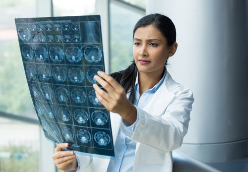
MRI and MRS are noninvasive imaging tests sensitive to changes occurring in dystrophic muscles that could be used in clinical trials.
While numerous studies support these magnetic resonance measures as high-quality Duchenne biomarkers, more robust information is needed to validate their use in trials.
A team led by researchers at the University of Florida used data from an ongoing natural history study (NCT01484678) that is following these two biomarkers in boys with DMD for up to 10 years.
For this specific study, a total of 160 patients (mean age, 8.6) were followed for up to 48 months or about four years.
The researchers used data from leg MRS and MRI scans to examine the progression of biomarker levels over time, as well as the ability of such markers to predict changes in walking ability and other physical skills.
Imaging scans of eight leg muscles were used to determine two main biomarkers — MRI T2, which can be used as a global measure of muscle health, and fat fraction, which is detected by MRS and quantifies the level of fat infiltration into muscle. Fat progressively accumulates and almost fully replaces muscle tissue in people with DMD.
Patients also underwent four functional tests of walking: the 10-minute walk/run, the 6-minute walk test, the four-stair climb test, and the supine-to-standing test.
All assessments were taken at the study’s start and again annually over the next four years. Of the 160 patients who entered the study, 79 completed the entire follow-up.
Fat fraction and MRI T2 levels of the vastus lateralis (VL) muscle — one of the muscles on the lateral, or side, of the thigh — as well as MRI T2 of the biceps femoris long head (BFLH; a muscle located on the back of the thigh), were the biomarkers with the greatest changes in patients and that worsened faster over time.
Most patients were able to walk unassisted at the beginning of the study, but more than 30% were no longer ambulatory four years later.
Looking at magnetic resonance biomarkers in relation to functional abilities, researchers found that a patient’s these values at study start predicted how well their physical skills improved, stabilized, or declined over the next 12 and 24 months. Higher magnetic resonance biomarker values correlated with greater loss of function.
Fat fraction and MRI T2 measures of the VL and soleus (the plantar flexor muscle of the ankle) muscles, and MRI T2 measures of the BFLH muscle were the strongest predictors of future loss of function, including losing the ability to walk, climb, or stand from a lying position.
The chances of remaining stable or improving over the next year were highest (over 50%) in patients with very low baseline VL fat fraction values (lower than 0.1), while the probability of declining or losing function was greatest for those with values above 0.4.
Patients with fast or slow disease progression by these magnetic resonance biomarkers tended to have a corresponding progression on functional tests.
“This study supports the strong relationship between lower extremity MR [ magnetic resonance] biomarkers and measures of clinical function, as well as the ability of MR biomarkers, particularly those from proximal muscles [those closest to the body], to predict future ambulatory function and important clinical milestones,” the researchers wrote.
“Most important, the hope is that inclusion of muscle MR biomarkers will accelerate development and approval of disease-modifying therapeutics for DMD,” they added.
Such markers could be a noninvasive way of evaluating muscle health, limiting the need for muscle biopsies, and addressing outcomes early, which would help reduce trial length or size, the team concluded.