
Pathway Key to Muscle Regeneration Doesn’t Work as It Should in Duchenne, Study Finds
Written by |
The Hippo signaling pathway — a key regulator of tissue development, and cell growth and death — does not work as it should in Duchenne muscular dystrophy and acts to prevent, rather than aid, the regeneration of muscle cells in DMD patients, a study reports.
A member of this pathway, a protein called YAP1, is an important regulator of muscle repair and regeneration after injury, and researchers suggest it could be a target for future Duchenne therapies. Interestingly, similar pathway dysregulation was not seen in other muscular dystrophies.
The study, “Hippo signaling pathway is altered in Duchenne muscular dystrophy” was published in the journal PLOS One.
The Hippo signaling pathway is a a very important controller of organ size, regulating the division rate of cells, cell death, and stem cells renewal. One of the players activated within the Hippo signaling pathway is the Yes-associated protein 1 or YAP1.
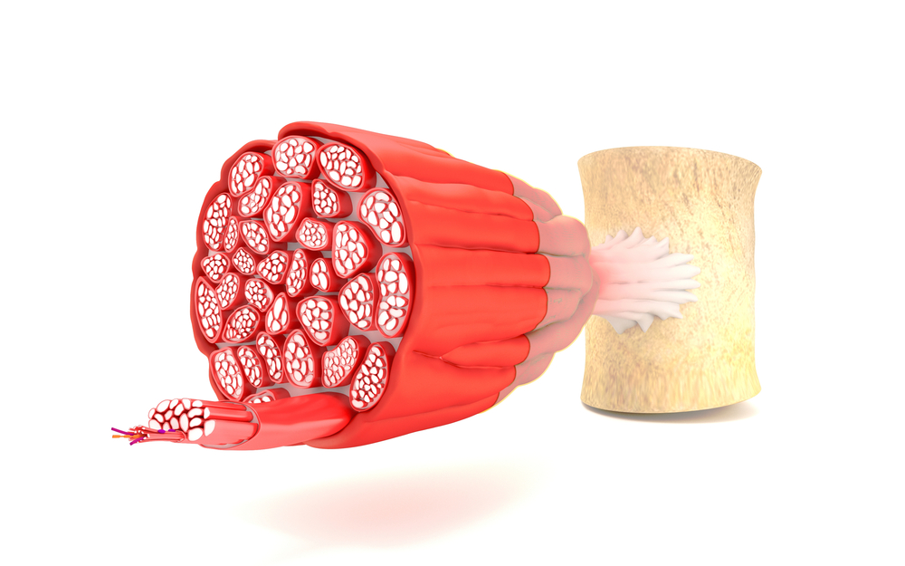
In skeletal muscle, or muscle responsible for voluntary movement, YAP is an important regulator of proliferation and other properties of myoblasts, the undifferentiated cells capable of giving rise to muscle cells.
Previous studies show that low levels, or a lower expression, of YAP is important in allowing myoblasts to transform and mature into muscle cells, while high levels of YAP keep myoblasts proliferating in their undifferentiated state.
A study analyzing gene expression in the muscle of a mouse model of DMD suggested that the Hippo pathway and, consequently, YAP were at increased expression.
“These results suggest that Hippo signaling may have an important but yet uncertain regulatory role in dystrophic skeletal muscle,” the researchers wrote.
However, few studies have analyzed YAP expression in DMD and other muscular dystrophies.
“The goal of the present study was to test the hypothesis that altered YAP signaling may contribute to dystrophic pathogenesis [disease development] in DMD muscle, becoming a pharmacological target of dystrophinopaties,” researchers added.
They analysed samples of the vastus lateralis, a muscle in the thigh, in five patients with DMD, ages 4 to 6. They also included samples from patients (five of each) with other muscular dystrophies — Becker muscular dystrophy (BMD), limb-girdle muscular dystrophy type 2A (LGMD2A), and limb-girdle muscular dystrophy type 2B (LGMD2B).
As controls, they used five muscle samples taken from healthy subjects, 3 to 50 years old.
Analysis showed that the production of YAP1 was reduced in the muscles of DMD patients, but not in the muscles of patients with other types of muscular dystrophy.
YAP1 levels can be reduced by the tumor suppressor 1/2 (LATS1/2) kinase. Researchers assessed the levels of LATS1/2 kinases and found that they were two times higher in Duchenne patients compared to healthy controls. Once again, and in agreement with the YAP1 expression, no increase was detected in children with other muscular dystrophies.
In the mdx mouse model, a widely used animal model for studying DMD, researchers also detected increased LATS1/2 kinase activity in five different muscles.
The levels of YAP1 varied, with some muscles showing no significant changes in YAP1 expression compared to healthy animals. In the gastrocnemious muscle, one of the two major muscles that make up the calf, they detected a 26 percent decrease in YAP1 expression, while its expression was slightly increased in muscles of the biceps and diaphragm (the thin skeletal muscle that separates the abdomen from the chest) — 5 and 16 percent, respectively.
They confirmed the expression of YAP1 by measuring of the levels of one of its targets, a factor called survivin, a protein that inhibits a form of programmed cell death called apoptosis.
Survivin levels were three times lower in the muscles from DMD patients compared to controls.
The known link between YAP1 and muscle regeneration/repair suggests that therapies targeting certain components of the Hipppo pathway “could become novel therapeutic targets for DMD treatment,” the study concluded.


Leave a comment
Fill in the required fields to post. Your email address will not be published.