New Potential Therapeutic Approach for Muscle Regeneration in Dysferlinopathy

A study recently published in the journal Skeletal Muscle revealed a new potential therapeutic approach to promote muscle regeneration in dysferlinopathy. The study is entitled “Upregulated IL-1β in dysferlin-deficient muscle attenuates regeneration by blunting the response to pro-inflammatory macrophages”, and was developed by researchers at the Children’s National Medical Center, the Kennedy Krieger Institute, the Johns Hopkins University School of Medicine and the University of Maryland.
Dysferlinopathy includes a group of rare, neuromuscular disorders caused by mutations in a gene called dysferlin (DYSF) and characterized by progressive muscle wasting. These mutations result in the production of a defective dysferlin protein, which plays a critical role in membrane repair and vesicle trafficking in the muscle tissue, leading to leakage of intracellular contents from the inside of muscle fibers. The disease primarily affects muscles of the limbs, the hips and shoulders, and individuals often become unable to walk.
Dysferlin-deficient muscles exhibit inflammatory foci and infiltration of specific immune cells called macrophages (a type of white blood cell) that ultimately leads to a decline in muscle function. In this context, macrophages are thought to inhibit the resolution of muscle regeneration. Two main classes of macrophages are known to play different roles during the acute injury process: the classical (M1) and the alternative (M2a).
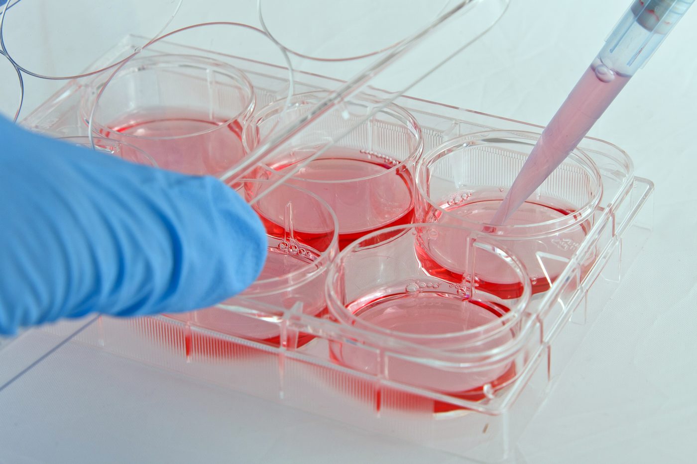
In the study, researchers investigated in more detail these two classes of macrophages and their individual role in chronic muscular disease. The team developed an in vitro co-culture system of macrophages together with healthy or dysferlin-deficient muscle cells. The co-cultures were analyzed for secreted factors and RNA expression (transcriptome).
Researchers found that dysferlin-deficient muscle had an excess of M1 macrophage markers in comparison to healthy muscle cells. In addition, dysferlin-deficient muscle poorly regenerated in response to injury by toxins. The team found that M1 macrophages inhibit muscle regeneration while M2a macrophages promote regeneration; this is especially observed in dysferlin-deficient muscle cells.
Regarding secreted factors and transcriptome analysis, two molecules were found to be relevant: interleukin (IL)-1 beta, which is secreted by M1 macrophages during acute injury, and IL-4 that is secreted by M2a macrophages. The team then treated muscle cells with IL-4 and found that the molecule improved muscle differentiation, while treatment with IL-1 beta inhibited muscle differentiation. Interestingly, blocking IL-1 beta signaling with a specific antibody led to a significant improvement in dysferlin-deficient muscle cell differentiation.
The authors concluded that M1 macrophages have an inhibitory effect on myogenesis (formation of muscular tissue), and that this effect is mediated by the IL-1 beta signaling pathway. The authors suggest that inhibition of the immune response mediated by M1 macrophages, including IL-1 beta inhibition, could be considered a potential therapeutic strategy to improve muscle regeneration in dysferlin-deficient disorders.


