
PPMD Webinar Takes In-depth Look at MD Cardiac Disease and Recent Research Efforts
Written by |
Magnetic Resonance Imaging (MRI) can catch signs of damage to the heart in muscular dystrophy (MD) patients earlier than traditionally-used echocardiograms, according to a webinar providing updates on the Parent Project for Muscular Dystrophy (PPMD)’s cardiac initiative.
The presentation took place Dec 12, which is almost halfway through the group’s monthlong fund-raising campaign for the projects, “The Heart of the Matter.” Throughout December an anonymous donor has agreed to match donations — doubling their impact — up to $275,000.
The webinar focused on three main projects from the initiative: understanding cardiac MRIs, the cardiac health of carrier moms, and the potential effects of gene therapy on the heart.
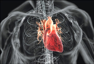
Imaging the DMD Heart
For the past decade, PPMD has spent $5 million on its heart initiative because all Duchenne muscular dystrophy (DMD) patients, and some of their female relatives, develop cardiomyopathy.
Kan Hor, MD, director of the cardiac MRI program at Nationwide Children’s Hospital in Columbus, Ohio, explained that new techniques for diagnosing and assessing cardiomyopathy (heart disease) are crucial for DMD patients. These patients are unlikely to complain of shortness of breath or other traditional symptoms of heart failure because they rarely are able to participate in physical activities that would lead to these symptoms.
Aside from patient complaints, doctors will use an ECG to determine if the heart is squeezing properly, and if it’s not, treatment will begin.
But Hor shows that MRI scans detect scarring on the heart in 84% of patients long before the damage affects the heart’s function. “If we are to impact the disease, we need to intervene before dysfunction, before too much scar formation,” said Hor.
The ongoing research is redefining the stages of cardiomyopathy for patients with muscular dystrophy and developing a model for disease progression. Most important, Hor says it’s clear that MRI scans should be routine in DMD patients to catch signs of heart disease as early as possible.
DMD Moms and Cardiomyopathy
May Ling Mah, MD, from Nationwide Children’s Hospital, presented the first year of data she has collected from what will be a 3-5 year study on the heart health of DMD mothers.
The first step of the study included genetically testing moms to see if they are carriers of the mutated dystrophin gene responsible for DMD. Fifty-seven moms in the study were confirmed carriers, 20 had sons with DMD but were not carriers, and another 25 controls did not have sons with DMD.
Previous studies have suggested that 9-15% of DMD moms between the ages of 31 and 50 experienced cardiac disease. But MRI scans showed that 44% of genetically-confirmed carrier moms in Mah’s study showed fibrosis compared to only one noncarrier mom, and none of the moms without DMD sons.
That number, despite being higher than the previously reported 15%, didn’t surprise Mah since previous studies hadn’t included genetic testing — lumping both carrier and non-carrier moms together. What did surprise her were the differences that showed up on the treadmill test, a noninvasive test in which participants walk or run on a treadmill while hooked up to an electrocardiogram to measure the stress the exercise puts on the heart.
Of the carrier moms, 25% had irregular heartbeats, specifically, premature ventricular contractions that occurred early during the recovery portion at the end of the workout. Mah explained that if a mom had an arrhythmia on the treadmill, there was a 69% chance she also had fibrosis, so the treadmill test could serve as a widely-available, noninvasive screen.
Still, while the carrier moms had fibrosis, their functional capacity was the same as noncarrier moms. “So your heart wasn’t really limiting you,” said Mah. But fibrosis on an MRI scan could be an early marker for cardiomyopathy in DMD moms, just like Hor suggests it should be for DMD boys.
Mah hopes to publish the data in the spring. In the meantime, the team is continuing to follow the moms and will be looking at the effects of age and stress in order to make definitive recommendations for screening and care.
Treating the DMD Heart with Gene Therapy
Since DMD is a genetic disease, it would fit that it could be treated by replacing the dysfunctional dystrophin gene with a functional one. The problem is that the gene is too large to fit into common viral vectors that could deliver it to a patient.
Several ongoing gene therapy studies are using micro-dystophin — a shorter version of the dystrophin gene — to slow disease progression and improve skeletal muscle in DMD patients, making their symptoms similar to those with Becker muscular dystrophy.
But these patients — and patients with genetic mutations that cause Becker MD — continue to develop cardiac symptoms. “This is what Becker patients have been telling us all along,” said Lee Sweeney, PhD, a professor at the University of Florida College of Medicine. Becker patients may have different genetic mutations that lead to less-severe skeletal muscle symptoms, but these mutations still can lead to severe cardiological symptoms.
Sweeney says researchers need to look to different gene therapy targets to treat the DMD heart than they are using to treat the skeletal muscles, such as calcium dysregulation, which animal studies have shown can lead to an overload of calcium straining the heart.
So, instead of delivering micro-dystrophin, Sweeney is using gene therapy in an attempt to correct calcium handling in the heart. If successful, the treatment could be used in combination with micro-dystrophin or as a monotherapy in Becker patients with heart disease.
His study hasn’t been published yet, but Sweeney did say his team has treated several animals and “I’ll tell you right now, their cardiac function looks normal at a time when it shouldn’t.”


Leave a comment
Fill in the required fields to post. Your email address will not be published.