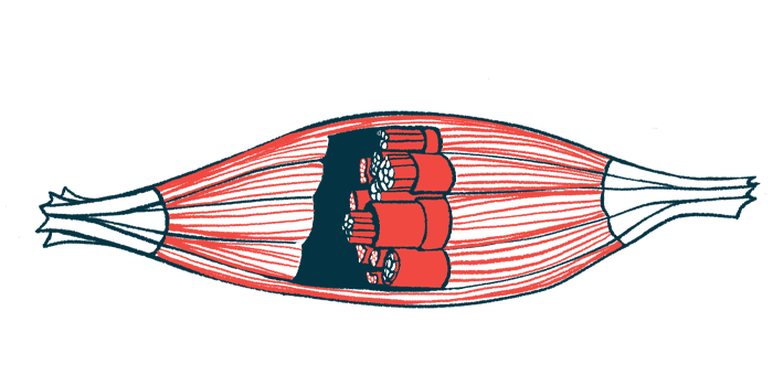
Restoring Piezo1 Ion Channel May Be Key to Repairing Muscle in DMD

An ion channel called Piezo1 regulates muscle stem cell (MuSC) states and the number of cells that respond quickly to muscle injury, promoting muscle repair, a study in mice shows.
Notably, this ion channel was found to be poorly activated in a mouse model of Duchenne muscular dystrophy (DMD), and increasing its activity restored MuSC state distribution and boosted muscle regeneration in this model.
“Even though progress has been made over the last decade on treatments for Duchenne muscular dystrophy, current strategies still do not take muscle stem cells into consideration,” Foteini Mourkioti, PhD, the study’s senior author and an assistant professor of orthopedic surgery at University of Pennsylvania’s Perelman School of Medicine, said in a press release.
“But if we focus on minimizing stem cell exhaustion and maintaining the regenerative ability of muscle stem cells, our work suggests that reactivating Piezo1 could be key to that and used alone or in combination with other therapies,” Mourkioti added.
The study, “Piezo1 regulates the regenerative capacity of skeletal muscles via orchestration of stem cell morphological states,” was published in the journal Science Advances.
Throughout life, adult muscle stem cells, also known as satellite cells, are essential for repairing and regenerating muscle tissue. They must replenish themselves, or “self-renew,” and they also develop into specialized cells that form new muscle fibers.
MuSCs are highly affected in DMD and other chronic muscle diseases, being unable to properly respond to muscle injury and leading to the accumulation of muscle damage.
How MuSCs sense their surroundings to know when and where there is muscle injury to act quickly, however, remains largely unknown.
A research team at Perelman discovered that MuSCs are divided into three functionally distinct populations: responsive cells, intermediate cells, and sensory cells that were closer to dormant stem cells.
By looking at the muscle environment of healthy mice in real time, the scientists first found that MuSCs showed different patterns of cellular extensions, or protrusions, that resembled axons — the cellular extensions used by nerve cells to communicate with each other.
MuSCs with fewer protrusions were in closer proximity to other cells, while those with more protrusions were rarely found near other cells and their protrusions extended throughout the muscle fiber.
“These data suggest that cells with fewer than three protrusions likely participate in cell-to-cell communication, while cells with four or more protrusions extend toward the muscle fiber, possibly ‘sensing’ the muscle environment,” the researchers wrote.
“This is in contrast to previous belief, which considered muscle stem cells to be simply round and dormant in undamaged muscles,” Mourkioti said.
Notably, upon muscle injury, MuSC protrusions were found to be shorter in length and the MuSC population shifted toward cells with fewer protrusions.
Cells with no or one protrusion were the first responders (responsive cells), self-renewing rapidly to repair muscle damage. Cells with four or more protrusions (sensory cells) “respond later, by losing their protrusions and transitioning toward intermediate and then responsive cells,” the researchers wrote.
These results highlight that “MuSC protrusions are not static structures but instead adapt to distinct regeneration phases, further supporting a coordinating role in muscle restoration,” they added.
Further analysis showed that this shift between different MuSC states was regulated by Piezo1, a mechanosensitive ion channel, meaning that it lets ions move in and out of the cells when subjected to mechanical stimuli.
Previous studies highlighted its involvement in mechanical signaling and cell fate regulation in bone-forming cells and axon regeneration. However, Piezo1’s role in MuSC regulation was unclear.
The team found that treating healthy mice with Yoda1, an activator of Piezo1, increased the number of responsive MuSCs and reduced the number of intermediate cells, showing that Piezo1’s activity primed MuSCs toward more responsive cells, even without muscle injury.
In agreement, deleting Piezo1 in MuSCs shifted their distribution toward less responsive cells, which was similar to what the team observed in a mouse model of DMD. These mice showed significantly lower Piezo1 levels in MuSCs, significantly longer protrusions, and significantly fewer responsive cells and more sensory cells relative to healthy mice.
Treating the DMD mouse model with Yoda1 was found to restore MuSC state distribution — with a significant increase in responsive cells — shorten protrusion length, and boost muscle regeneration.
These findings “advance our fundamental understanding of how stem cells respond to injury and identify Piezo1 as a key regulator for adjusting stem cell states essential for regeneration,” the scientists wrote.
“We suggest that under the high regenerative demands in the diseased muscles, dystrophic [MuSCs] are unable to channel the information in a timely manner, leading to the reported improper MuSC activation in dystrophic muscles,” they added.
Further studies are needed to better understand Piezo1’s role in muscle regeneration and its potential as a therapeutic target in larger animal models of DMD. Such information may also be relevant for other muscle conditions resulting from compromised MuSC abilities, including from natural aging.
“Understanding the way that Piezo1 works better could be useful in designing more accurate therapies,” Mourkioti said.


