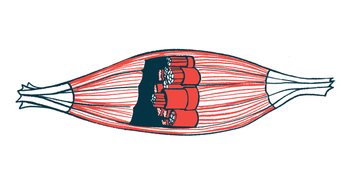
New 3D muscle model will help scientists study therapies for DMD
Created from patient cells, model shows damage seen in Duchenne

Researchers have created a three-dimensional (3D) artificial muscle model, derived from patient cells, that accurately replicates the damage observed in people with Duchenne muscular dystrophy (DMD).
This new model, the scientists believe, will enable more efficient testing of treatments that could potentially reverse such muscle damage.
The findings were described in “Mimicking sarcolemmal damage in vitro: a contractile 3D model of skeletal muscle for drug testing in Duchenne muscular dystrophy,” a study published in the journal Biofabrication. The team already is planning to develop a more advanced version of the 3D muscle model, incorporating additional technologies that could expedite testing of potential therapies.
“It is a source of pride to know that we have funded a project that can expedite the research process through the creation of an artificial muscle-on-a-chip,” Silvia Ávila, president of the Duchenne Parent Project Spain, which funded the study, said in a press release.
“On one hand, we reduce the use of animals in the laboratory, and on the other hand, we shorten the time to turn a potential therapy into reality. This is crucial for a disease that is robbing our children of time and quality of life,” Ávila added.
Scientists aimed to model primary cause of DMD for first time
In Duchenne, the most common type of muscular dystrophy, mutations in the DMD gene cause a lack of production of dystrophin, a protein that helps protect muscle cells from wear and tear. As such, patients experience progressive muscle wasting.
Particularly, a lack of dystrophin causes damage to the sarcolemma — the muscle cell membrane — resulting in damage to muscle fibers when they contract.
One challenge in developing therapeutics for DMD is the lack of preclinical models that accurately reflect the complexity of the disease.
3D models derived from patient cells have emerged as a promising approach for more accurately modeling the human condition. These artificial — but functional — tissues will have the same cellular characteristics as the patient from which they were derived.
Now, using cells from DMD patients, a team of scientists successfully developed a 3D artificial muscle model for Duchenne. The engineered muscles were functional, with the ability to contract in response to electrical stimulation.
Importantly, prolonged muscle contractions led to sarcolemmal damage, mirroring the loss of muscle fiber integrity typically observed in DMD patients. Meanwhile, such damage wasn’t seen in muscles derived from healthy cells.
“We worked for a long time on different protocols until we managed to make that damage appear in the cells of patients, but not in the control cells of people without Duchenne,” said Ainoa Tejedera-Villafranca, a doctoral student at the Institute for Bioengineering of Catalonia (IBEC), in Spain, and the study’s first author.
“It’s delicate because if you stimulate the muscle, you can also cause fiber breakage in healthy cells, just as it happens when we play sports and experience muscle soreness,” Tejedera-Villafranca added.
We worked for a long time on different protocols until we managed to make [muscle fiber] damage appear in the cells of patients, but not in the control cells of people without Duchenne.
After the damage was induced, DMD muscle tissues showed a tendency to fatigue with repeated stimulation, indicating that sarcolemmal damage compromises muscle function as it does in the human condition.
“The novelty of this study lies in our attempt to model the primary cause of the disease, which is damage to the sarcolemma,” said Juanma M. Fernández-Costa, PhD, a senior researcher at IBEC and the study’s senior author.
“It was crucial for us to replicate this in the laboratory, and we have successfully accomplished it,” Fernández-Costa said, adding, “This had not been done previously.”
New 3D muscle model already put to use in lab
Now, the researchers hope the model can be used to identify potential treatments to reverse DMD-associated muscle damage. To date, most therapies for Duchenne can only ease symptoms.
This possibility already has been evaluated by the team, which treated the muscle cells with therapies to increase levels of utrophin, a protein with a similar structure and function to dystrophin — which has been identified as a possible DMD treatment target.
The small molecules halofuginone or ezutromid were found to have the opposite of the intended effect, however. Specifically, muscle contractile function was notably reduced. Moreover, halofuginone caused muscle atrophy, or wasting.
According to the researchers, these findings were consistent with those of previous Phase 2 trials of the therapies, which failed to show efficacy.
Another investigational utrophin upregulator, from SOM Innovation Biotech, was found to increase muscle contractions, but did not protect against sarcolemmal damage.
“Altogether, these findings underscore the potential of our DMD model to serve as a platform to gain a deeper understanding of how potential drugs and other molecules can influence skeletal muscle functionality,” the researchers wrote.
Now, the scientists are working on an enhanced “organ-on-a-chip” model that will incorporate sensors and other technologies to enable better monitoring of cell damage, further accelerating the testing of therapeutic molecules.
The team is continuing to work with the parents of DMD children through the Duchenne Parent Project Spain, the nonprofit that is funding this research.
Fernández-Costa said the collaboration “is a fantastic example of how researchers can work together with patients and their families to pursue common goals.”


