Researchers Engineer Skin Cells from Duchenne MD Patients, Providing New Understanding and Therapeutic Avenues
Written by |

Researchers at Johns Hopkins Medicine in Baltimore were able to generate muscle cells from skin cells of Duchenne muscular dystrophy (DMD) patients, forming human cell models of different DMD genetic mutations that will allow researchers to further understand the disease and test new therapies. The scientists were also able to correct abnormal levels of protein expression and lead the cells to more normal “muscle-like” behavior through genetic manipulation and pharmacological therapy.
The research paper, “Concordant but Varied Phenotypes among Duchenne Muscular Dystrophy Patient-Specific Myoblasts Derived using a Human iPSC-Based Model,” was published in Cell Reports.
Duchenne muscular dystrophy (DMD), one of nine types of muscular dystrophy, is an X chromosome-linked genetic disorder where a mutation leads to the absence of the cytoskeletal protein dystrophin, resulting in progressive degeneration, muscle weakness and deterioration, and cardiac dysfunction. DMD primarily affects boys and is associated with poor survival outcomes. There is currently no cure for DMD and no approved drug in the U.S. to treat this condition. Although there are several animal models of DMD for research, there are no human cell models that carry specific mutations in the dystrophin gene — a model that would greatly help understand the disease and help develop new therapies.
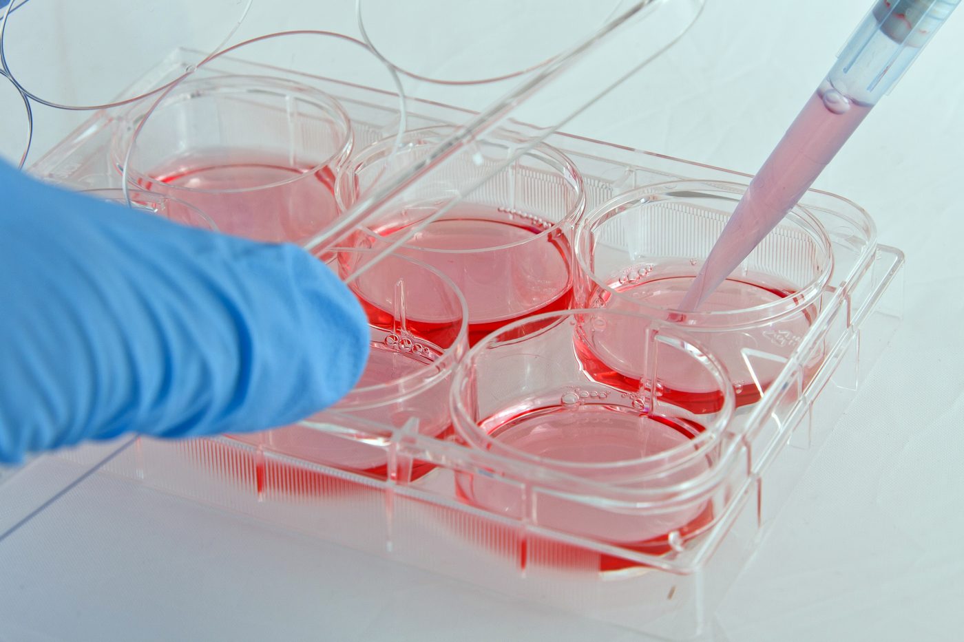
The team developed a chemical compound recipe that successfully induces DMD in human induced pluripotent stem cells (hiPSCs). In vitro culture with the compound CHIR99021, followed by treatment with the compound DAPT, caused cells to spontaneously contract and behave as muscle fibers, which are formed when myoblasts mature and fuse, forming myotubes that then mature into muscle fibers.
“The fusion of myoblasts into myotubes is one of the most distinctive features of skeletal muscle,” study leader Dr. Gabsang Lee, an assistant professor of neurology at Johns Hopkins School of Medicine, explained in a news release. “What we can now do is make fusion-capable myoblasts, which are easy to grow and therefore make it easier to study how diseases like DMD interfere with the fusion process.”
The researchers applied this process in the development of the human model of DMD by harvesting skin cells from DMD patients with different genetic mutations and healthy donors. From both healthy iPS cells and DMD iPS cells, the scientists were able to create myoblasts that could generate myotubes. DMD-derived cells were, however, much less efficient at the fusion step and were not able to contract.
The researchers then analyzed gene and protein activity to look for common aspects between the mutations. Compared to healthy cells, DMD myoblasts did not make dystrophin and showed greater activity in genes associated with wound healing and inflammation. Specifically, myoblasts generated from the DMD patients showed increased activity in the genes BMP4 and TGFβ.
The researchers were then able to correct DMD mutations in the laboratory by introducing a small artificial chromosome carrying a good copy of the dystrophin gene in the recovered skin cells from patients, reprogramming them to become iPS cells and growing them into myoblasts, which were able to fuse with the wounded muscle in mice. The scientists also obtained normal fusion levels by treating cells with the compounds LDN193189 and SB431542, inhibitors of the overactive genes BMP and TGFβ.
Even though these are early-stage data, the results might account for the first steps into a new therapy that will allow scientists to genetically correct the mutations that lead to the devastating disease.


